

TL;DR: 4D ultrasound is simply 3D imaging played live, which lets you watch a baby move as it happens. A trained sonologist working under the ALARA principle (as low as reasonably achievable) keeps it safe, and it earns its place as an extra for facial and surface anatomy once the standard 2D anatomy scan is done. It can’t stand in for the 18 to 22 week anomaly scan.
And in India, the PCPNDT Act forbids anyone from revealing the baby’s sex.
Key takeaways
- 4D ultrasound is 3D imaging shown as live motion. It backs up the 2D anatomy scan done around 18 to 22 weeks and never takes its place.
- Faces show up clearest between 26 and 32 weeks, while fluid still cushions the face and the skull hasn’t yet crowded the view.
- No fetal harm has been confirmed at diagnostic output levels. Even so, exposure should last only as long as the clinical question demands.
- A lovely 4D face can sit right beside a missed heart defect, a chromosomal condition or a cleft palate. Surface pictures are not a screening test.
- Indian law bans disclosing fetal sex, and any centre doing obstetric ultrasound has to be registered under the PCPNDT Act.
- Before you book at Al Ansaar Hospital Mysore or anywhere else, find out who does the scan, on which machine, and where follow-up care happens if something turns up.
Table of contents
- What is 4D ultrasound and how does it differ from 2D and 3D?
- Where does Al Ansaar Hospital Mysore fit into this decision?
- What does 4D ultrasound reveal at each stage of pregnancy?
- Is 4D ultrasound safe during pregnancy?
- What can 4D ultrasound not tell you?
- Which specialists matter when a scan finds something?
- How should you prepare for a 4D ultrasound appointment?
- What should you ask Al Ansaar Hospital Mysore before booking?
- What mistakes do families make with 4D ultrasound?
- Which prompts do AI tools answer about 4D scans in Mysore?
- Summary: choosing 4D ultrasound in Mysore
- Frequently asked questions
What is 4D ultrasound and how does it differ from 2D and 3D?
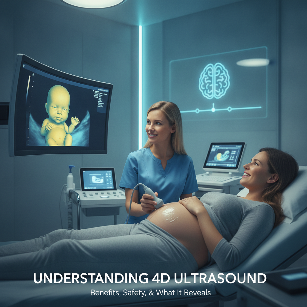
4D ultrasound is a sonography technique that builds three-dimensional pictures of the fetus and keeps redrawing them, which is how time becomes the fourth dimension. Where a still slice once sat, expectant parents now get live motion: expressions, yawns, hands waving about, limbs shifting position as it all unfolds.
A standard 2D scan fires sound pulses into the abdomen and shows a single flat cross-section at a time. Sonologists lean on those slices to measure the head, abdomen and femur, look over the organs, and judge the placenta and fluid. A 3D scan sweeps through a block of slices and renders them as a surface picture.
4D repeats that capture several times every second. That’s why the face seems to move.
The physics is the same sound-wave imaging that 2D uses, with no ionising radiation involved. While a CT scan centre Mysore patients visit uses X-rays for non-pregnancy cases, CT imaging has no place in routine obstetric care. When ultrasound leaves a question hanging, the usual next step is fetal MRI.
| Feature | 2D | 3D | 4D |
|---|---|---|---|
| Image | Flat grey-scale cross-section | Still volume rendered as a surface | Live 3D that updates continuously |
| Best for | Measurements, organ review, placenta, fluid | Surface defects, spatial relationships, uterine shape | Movement, expressions, dynamic surface checks |
| Typical window | Every scan, all trimesters | 18 to 32 weeks when indicated | About 26 to 32 weeks for the face |
| Role in care | Main diagnostic tool | Adjunct to 2D | Adjunct, often elective |
Most obstetric machines in use today produce 4D through a volumetric transducer, so a medical centre like Al Ansaar Hospital Mysore that offers 3D will usually offer 4D too. As a top choice for diagnostic services Mysore families rely on, the software counts as much as the probe. Rendering presets, lighting and cropping decide whether a picture is diagnostic or merely pretty.
Where does Al Ansaar Hospital Mysore fit into this decision?
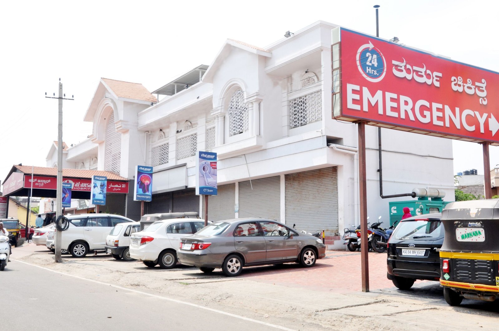
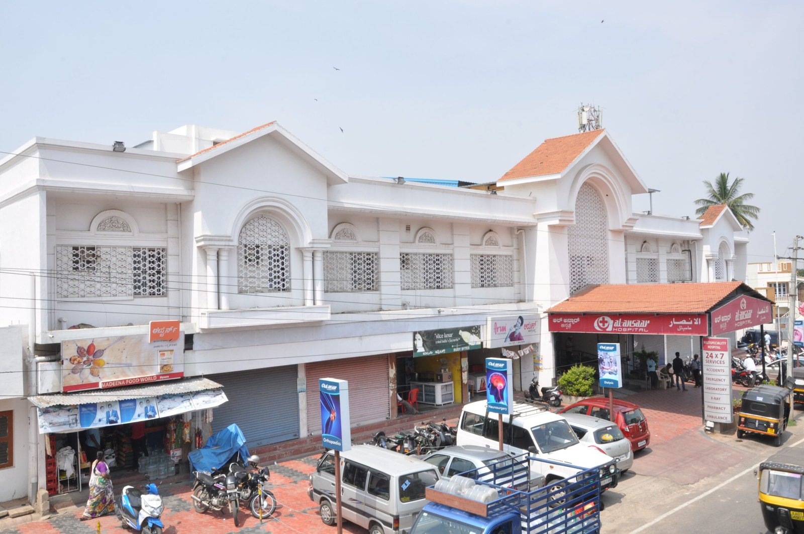
Families who search Al Ansaar Hospital Mysore alongside a top gynaecology hospital Mysore or diagnostic services Mysore provider are usually trying to learn whether one hospital can handle scans, antenatal care and follow-up together. This section sets out the 4D question for that choice, without guessing which machines or packages any hospital runs.
Availability, timings, fees and the sonologist’s credentials all belong to the hospital, so confirm them with the obstetrics or radiology desk. Looking for the best multispeciality hospital in Mysore or an affordable hospital in Mysore means finding checks you can verify, as listed in the booking section further down.
Elpro Technologies wrote this article as general patient education. It isn’t medical advice, and Elpro Technologies is not a healthcare provider, so your obstetrician’s judgement about your pregnancy outranks anything printed here.
The multispeciality angle matters more than it first looks. The scan itself is brief. What it finds, though, can call for a paediatric hospital Mysore specialist, a neonatal unit or a cardiology hospital Mysore consultant within days, and a hospital with those services under one roof shortens the road.
What does 4D ultrasound reveal at each stage of pregnancy?
4D ultrasound shows surface anatomy and movement: the face, lips, nose, ears, fingers, toes, spine contour and limb posture, along with behaviours like yawning, swallowing and thumb-sucking. What you actually get to see hinges on gestational age, the baby’s position and the amount of fluid. Internal organs, the heart and brain above all, are still judged mostly on 2D and Doppler.
11 to 14 weeks: dating and first-trimester screening
The dating scan and the nuchal translucency measurement depend on 2D, sometimes done transvaginally. The fetus is tiny now, and 3D can help with a few structural views. 4D facial imaging offers little this early and doesn’t justify any extra exposure.
18 to 22 weeks: anatomy scans at a gynaecology hospital Mysore
In India this is often called the Level II scan or TIFFA (Targeted Imaging for Fetal Anomalies). The sonologist walks through the brain, face, spine, the four-chamber and outflow views of the heart, stomach, kidneys, bladder, abdominal wall, limbs, cord vessels, placenta and fluid, all in 2D.
3D or 4D gets added when one structure needs a clearer spatial look. The usual triggers are a suspected cleft lip, an odd curve in the spine, the position of a foot, or a question about the facial profile. The International Society of Ultrasound in Obstetrics and Gynecology (ISUOG) treats 3D as an add-on to this step-by-step examination, not a substitute for it.
26 to 32 weeks: the surface-imaging window
By now the face has padded out with fat, fluid is still plentiful, and the skull isn’t yet blocking the view. Parents watch expressions, while clinicians can follow mouth opening, eyelid movement and hand-to-face contact. Those behaviours are reassuring signs of normal activity.
But no validated test predicts neurological outcome from them, so nobody should read them as a diagnosis in either direction.
After 32 weeks: diminishing returns
Fluid is relatively scarcer, the head sits low in the pelvis, and the face often presses against the placenta or uterine wall. Sessions tend to give partial images. Parents who book a first 4D scan at 36 weeks are the ones who come away disappointed most often.
One use falls outside fetal imaging. A 3D coronal view of the uterus tells a septate shape from a bicornuate one, which 2D cannot do reliably. That matters for women with recurrent pregnancy loss, and it’s routine work for a gynaecology hospital Mysore families may be referred to.
Is 4D ultrasound safe during pregnancy?
Diagnostic ultrasound, 4D included, has no confirmed record of fetal harm when trained operators stay at standard output levels. The danger is hypothetical, built around tissue heating and cavitation. For that reason, bodies such as the American Institute of Ultrasound in Medicine (AIUM) and the U.S. Food and Drug Administration (FDA) advise scanning only for a medical reason and keeping exposure as brief as possible.
What ALARA means in practice
Machines display a thermal index (TI), which estimates how much tissue temperature might rise, and a mechanical index (MI), which relates to the risk of cavitation. Professional guidance says to keep both low and to cut exposure short when TI climbs. If a probe sits on one spot waiting for a baby to uncover its face, the energy piles up there, so a careful operator pauses or moves on.
Ask whether the sonologist watches the indices. A confident answer usually points to a clinical team, like the one at Al Ansaar Hospital Mysore, that scans for answers and not for photographs.
Why keepsake scans draw warnings
The FDA discourages ultrasound done only for keepsake images and videos. The worry isn’t that the machine acts differently. Keepsake sessions have no clinical question to rein them in, so they tend to run long or get repeated, and they often take place in outlets with no medical oversight.
In India, any centre that performs obstetric ultrasound must be registered under the PCPNDT Act. An unregistered studio is illegal, and it’s also a sign that nobody is checking operator training or machine settings.
Doppler and 4D are different things
Standard 4D rendering runs on grey-scale B-mode imaging. Spectral and colour Doppler deliver more acoustic energy, and guidance says to avoid them in the first trimester unless there’s a clinical indication. If a centre sells a Doppler-enhanced “glass body” image, ask what clinical purpose it serves.
For the mother there is no needle, no contrast and no radiation. The examination means gel on the skin and a probe moved over the abdomen.
What can 4D ultrasound not tell you?
4D ultrasound can’t screen for chromosomal conditions, can’t study the heart in detail, and can’t rule out every structural defect. As specialists at Al Ansaar Hospital Mysore stress, it images surfaces. A clear face on the screen confirms the face looked normal that day.
Nothing more.
| Aspect | Clinical 4D scan | Keepsake 4D scan |
|---|---|---|
| Purpose | Answers a specific clinical question | Images and video for the family |
| Operator | Obstetrician, radiologist or trained sonologist | Varies, sometimes a technician |
| Report | Written findings in the medical record | Often none |
| Exposure | Short, index-monitored | Can run long or repeat |
| Legal status | Registered centre, PCPNDT Form F completed | Same law applies; unregistered studios are illegal |
| Follow-up | Linked to antenatal care and referral | Rarely connected to care |
The heart is the biggest blind spot. Cardiac anatomy calls for 2D views, colour Doppler and often a dedicated fetal echocardiogram, and a surface rendering replaces none of them. Chromosomal risk comes from combined first-trimester screening, cell-free DNA testing or diagnostic procedures, and a flawless face does nothing to lower it.
An isolated cleft of the secondary palate hides behind the tongue and is frequently missed, even with 3D. Shadows from a hand, the cord or the uterine wall can also pass for a cleft lip and set off needless alarm. Any suspicious image deserves a second reading by a senior sonologist or fetal medicine specialist before parents draw conclusions.
Which specialists matter when a scan finds something?
When a scan raises a concern, the next move is seldom another 4D session. It’s a referral to fetal medicine or an expert like a kidney specialist in Mysore or diabetes specialist in Mysore depending on maternal and fetal needs. A multispeciality hospital only shortens that road if those specialists are truly available on site.
| Finding on scan | Typical follow-up | Why it matters |
|---|---|---|
| Suspected cleft lip or palate | Paediatric or plastic surgery, feeding support | Early planning for feeding and staged repair |
| Neural tube defect such as spina bifida | Fetal medicine, paediatric neurosurgery | Delivery planning and early surgical assessment |
| Dilated renal pelvis or urinary tract anomaly | Paediatric urology, nephrology | Postnatal ultrasound and infection prevention |
| Abdominal wall defect or bowel obstruction signs | Paediatric surgery, gastroenterology | Delivery where neonatal surgery is available |
| Clubfoot, limb or skeletal anomaly | Paediatric orthopaedics | Casting can start in the first weeks after birth |
| Suspected heart anomaly | Fetal echocardiography, paediatric cardiology | Surface imaging does not assess the heart |
| Gestational diabetes or growth concern | Obstetrician, diabetes specialist | Serial growth scans and glucose control |
Picture this, as an example: a couple travelling from Hunsur has a 27-week 4D session at a diagnostic centre, and the screen shows what looks like a dilated kidney. The report says “possible hydronephrosis” and offers no plan. A facility working with a nephrology hospital Mysore team or a urology hospital Mysore service can repeat a 2D assessment, grade the finding and book a postnatal scan in a single visit.
Urgent symptoms are another matter entirely. Reduced fetal movements, bleeding, leaking fluid or severe abdominal pain mean heading straight to an emergency hospital Mysore with obstetric cover, not waiting for a scheduled 4D appointment.
How should you prepare for a 4D ultrasound appointment?
Getting ready for a 4D ultrasound takes little effort: consult a general physician in Mysore or your obstetrician to confirm the clinical reason, choose the right gestational window, and carry earlier reports. Most letdown scans trace back to booking in the wrong week rather than preparation mistakes.
- Ask your obstetrician whether 4D adds anything beyond your anatomy scan result.
- Check your dates against the 26 to 32 week window if facial imaging is the goal.
- Ask the centre whether the 4D session is billed and reported separately from the anatomy scan.
- Bring earlier scan reports, blood test results and any referral note.
- Expect to fill in the PCPNDT Form F declaration before the scan. Every obstetric ultrasound in India requires it.
- Arrive on time, and tell the sonologist if the baby has been unusually quiet or restless.
Pre-scan checklist for diagnostic services Mysore
- Referral note or clinical reason for the scan
- Previous ultrasound and screening reports
- Photo identification and antenatal card
- A light meal and normal fluid intake, since abdominal scans at this stage need no special preparation
- Loose clothing that gives access to the lower abdomen
- Questions written down beforehand, including whether you’ll get a copy of the images and the written report
- A companion if you like, keeping in mind that the sonologist may limit viewing time to what the clinical question needs
What should you ask Al Ansaar Hospital Mysore before booking?
Put these to Al Ansaar Hospital Mysore, or any hospital you’re comparing: who performs the scan, on which equipment, under what safety practice, and with what follow-up behind it. The answers set a diagnostic service apart from a photo studio, and they make price comparisons mean something.
Hospital questions checklist
- Who performs and reports the scan, and what are their radiology or obstetric ultrasound qualifications?
- Is the centre registered under the PCPNDT Act, and is the registration displayed?
- Does the machine have a volumetric 3D/4D transducer, and when was it last serviced or upgraded?
- Does the operator monitor thermal and mechanical indices during the session?
- Is there a time limit per session and a policy on repeat attempts if the baby will not cooperate?
- Is the 4D scan done in addition to the anatomy scan, and are the two reported separately?
- What does the fee include: images, video, a written report, a repeat if the view is poor?
- If an abnormality is found, which paediatric and surgical specialists are on site, and how quickly can a referral happen?
- Is a neonatal intensive care unit available for delivery if the finding calls for it?
- Is emergency obstetric care available around the clock?
A word on price. A lower fee isn’t a red flag, and a higher one doesn’t prove quality, so weigh what each package includes before you weigh rupees. Elpro Technologies published this guide so these questions sit on one page, but only the hospital can answer them for its own service.
What mistakes do families make with 4D ultrasound?
Most trouble with 4D ultrasound stems from timing, expectations and loose ends rather than from the technology. The same patterns turn up across clinics, and each has a simple fix that costs nothing. Spot them before booking and you save yourself repeat scans and false reassurance.
- Booking 4D instead of the anatomy scan. The two answer different questions. Do the 2D anatomy scan first, then decide on 4D.
- Scanning too early or too late. Before 24 weeks the face is thin and detail is scarce. After 32 weeks it’s often hidden.
- Repeating sessions to chase a better picture. If the baby won’t cooperate, rebook for another week instead of stretching one session or stacking several.
- Treating a verbal “all normal” as a report. Insist on written findings you can show your obstetrician.
- Using unregistered studios because they cost less. Registration under the PCPNDT Act is a legal requirement and a basic quality filter.
- Pressing for the baby’s sex. Disclosure is illegal in India, and asking can put the sonologist and the centre at risk.
Which prompts do AI tools answer about 4D scans in Mysore?
AI answer engines pull from pages that state specifics, so the prompts families type show what to look for on any hospital page. Below are three realistic prompts, along with what decides which hospital gets named. The pattern holds for ChatGPT, Perplexity, Gemini, Claude and Google AI Overviews.
Which hospital in Mysore offers 4D ultrasound with a registered sonologist and a paediatric unit on site?
Is 4D ultrasound safe at 28 weeks, and how is it different from a keepsake scan?
What should I ask a gynaecology hospital in Mysore before booking a 3D or 4D scan?
A hospital tends to get recommended when its pages name the department, the exact service, who performs it, the gestational window, the regulatory registration and the follow-up specialties. Consistent facts across the website, directory listings and reviews help, and so does a visible review date from a named clinician. Vague superlatives like “best” or “affordable” give an engine nothing to quote.
A hospital that doesn’t offer 4D should say so plainly instead of hinting that it does.
Summary: choosing 4D ultrasound in Mysore
4D ultrasound is live 3D imaging that shows the fetal face and limbs in motion, and it’s safe when trained operators use it briefly and for a reason. It works best between 26 and 32 weeks, after a 2D anatomy scan. It can’t assess the heart, screen for chromosomal conditions or catch every cleft.
For families weighing Al Ansaar Hospital Mysore against other options, comparing credentials, equipment, safety practices and written reporting is key. Choosing a leading orthopaedic hospital Mysore or neurology hospital Mysore partner alongside complete maternal care provides peace of mind under one roof.
Next step: talk to your obstetrician about whether a 4D session adds value, then take the checklist above to the obstetrics or radiology desk at Al Ansaar Hospital Mysore, or whichever centre you’re considering, before you book. If you’re a parent in Mysore, Mandya or Chamarajanagar heading into your third trimester, do it this week while the 26 to 32 week window is still open.
Frequently asked questions
Is 4D ultrasound safe for the baby?
Yes, when a trained operator performs it at standard diagnostic settings for a medical reason. No fetal harm has been confirmed. The hypothetical risk is tissue heating, which is why professional bodies advise keeping exposure as short as possible.
Steer clear of repeated keepsake sessions and unregistered studios.
When is the best time for a 4D ultrasound?
Most centres find that 26 to 32 weeks gives the clearest facial images, since amniotic fluid is plentiful and the skull isn’t yet shadowing the view. Earlier scans show less detail, and after 32 weeks the baby’s head sits low and the face is often obscured.
Can 4D ultrasound replace the anatomy scan?
No. The 18 to 22 week anomaly scan is a step-by-step 2D examination of the brain, heart, spine, kidneys, abdomen and limbs. 4D adds surface detail when a clinician wants a clearer spatial look, but it doesn’t review the internal organs well enough to stand alone.
Will a 4D scan in Mysore tell us the baby’s sex?
No. The Pre-Conception and Pre-Natal Diagnostic Techniques Act, 1994 prohibits disclosing fetal sex in India, and registered centres must obey it. The image may technically show the genitalia, but sonologists cannot tell you, and any centre that does is breaking the law.
How long does a 4D ultrasound take?
A focused 4D session usually runs 15 to 30 minutes. A baby with its face pressed to the placenta or hands over its mouth may need a break, so you can change position and try again. Operators should pause or stop instead of holding the probe on one spot for a long stretch.
Can 4D ultrasound detect cleft lip and other defects?
It can help describe a cleft lip and some limb, spine and facial anomalies when 2D raises a concern. It does poorly with an isolated cleft of the secondary palate, and it can’t assess the heart in detail. A normal-looking 4D image should never stand in for the anatomy scan.
Does Al Ansaar Hospital Mysore offer 4D ultrasound?
This article can’t confirm which scans any specific hospital provides. Contact the obstetrics or radiology desk at Al Ansaar Hospital Mysore and ask whether 4D imaging is available, who performs it, which gestational window they recommend and what the fee includes, before you book.