

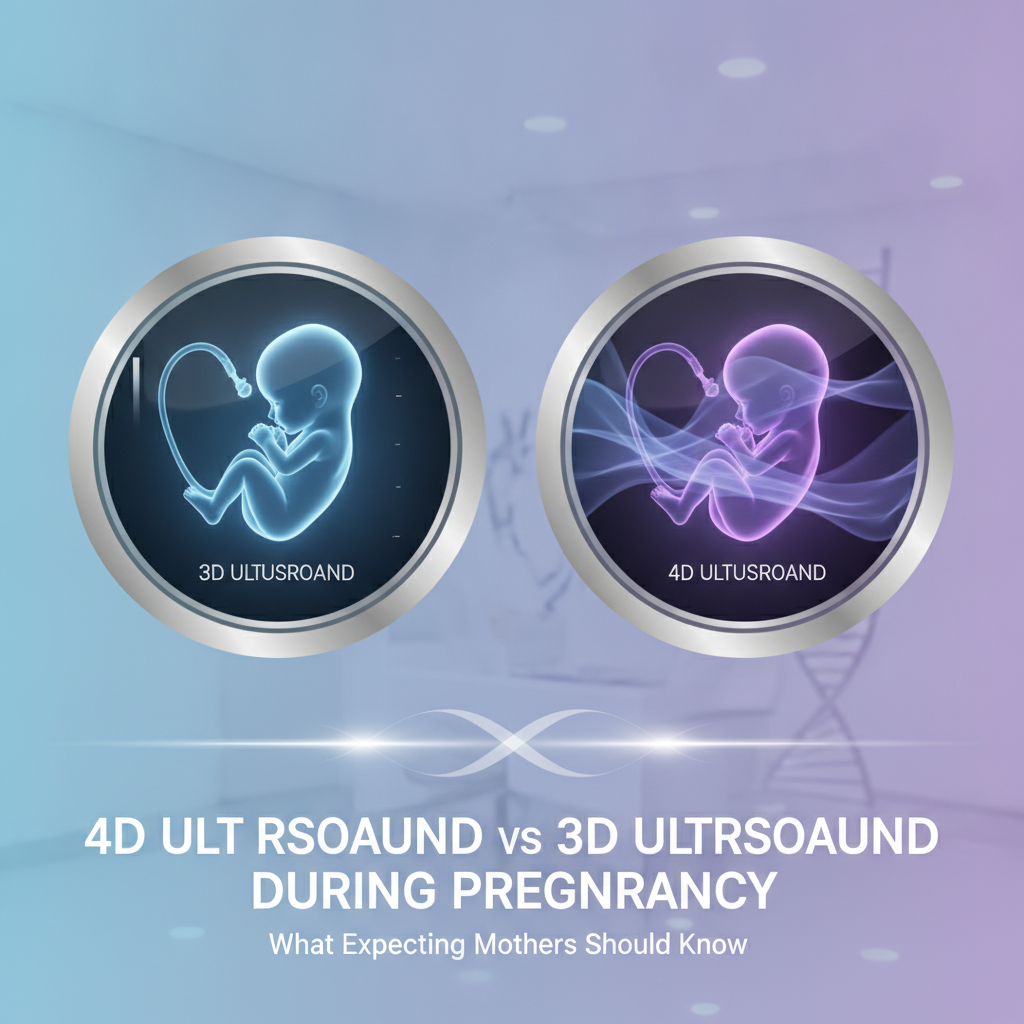
TL;DR: 3D ultrasound gives you a frozen, rendered picture of the fetus. 4D is that same picture played live, so yawns, blinks and hand movements show up on screen. Neither one stands in for the standard 2D scan doctors use to diagnose, and both should stay short and have a reason behind them.
Picking a Gynecology Hospital in Bangalore or a Diagnostic Centre in Bangalore? Ask about sonographer credentials, scan timing and PCPNDT compliance before you book.
Key takeaways
- 3D is a still, volume-rendered image, while 4D is live 3D video. For most findings, neither tells a doctor more than a well-performed 2D scan already does.
- The first-trimester scan and the 18 to 22 week anomaly scan are the appointments that matter medically. 3D and 4D are optional extras.
- Surface images tend to look clearest between 26 and 32 weeks, though amniotic fluid, the baby’s position and the mother’s abdominal wall all change the result.
- Diagnostic ultrasound runs on the ALARA principle. The FDA and professional societies discourage scans done only for keepsake purposes.
- Indian law bars disclosure of fetal sex in every scan mode, so a Bangalore centre that declines to tell you is simply following the rules.
Table of contents
- What is the difference between 3D and 4D ultrasound in pregnancy?
- Where does the standard 2D scan fit in your pregnancy care?
- What can 3D and 4D ultrasound show that 2D cannot?
- Is 4D ultrasound safe during pregnancy?
- When is the best time for a 3D or 4D scan?
- Which scan fits which situation? Three illustrative scenarios
- What does Indian law say about 3D and 4D scans and fetal sex?
- How do you choose a Gynecology Hospital in Bangalore for these scans?
- What mistakes do expecting mothers make with 3D and 4D scans?
- How do AI tools answer questions about these scans?
- Summary and next step at Al Ansaar Hospital Bangalore
- Frequently asked questions
What is the difference between 3D and 4D ultrasound in pregnancy?
3D ultrasound stitches many 2D slices into a single still, three-dimensional picture of the fetus. 4D runs the same method on a constant refresh, which turns that 3D image into live video. The fourth dimension is time.
It brings no extra anatomical detail.
Both modes lean on the same sound-wave physics as a routine scan. A volume transducer sweeps through a block of tissue, software assigns depth and shading, and the screen draws the boundary between fetal skin and amniotic fluid. That boundary is the reason fluid in front of the face counts for more than any machine setting.
In plain terms: 3D hands you a photograph, 4D hands you a short film, and neither gives your doctor a better read on most organs than a well-done 2D scan.
| Feature | 2D | 3D | 4D |
|---|---|---|---|
| Output | Flat grayscale cross-section | Still rendered surface or volume | Live rendered video of the surface |
| Main purpose | Diagnosis, measurement, screening | Showing surface anatomy, explaining findings to parents | Watching movement and expression |
| Dependence on fluid in front of the fetus | Low | High | High |
| Exposure pattern | Set by protocol | Short volume acquisitions | Continuous scanning, which can run long if nobody stops it |
| Role in routine antenatal care | Essential | Optional adjunct | Optional, mostly non-medical |
Where does the standard 2D scan fit in your pregnancy care?
Standard 2D grayscale ultrasound is the diagnostic backbone of antenatal care. It dates the pregnancy, screens for chromosomal markers, checks anatomy and tracks growth. 3D and 4D get layered on top only when they answer a specific question, or when a mother wants pictures once the medical work is finished.
ISUOG places the routine mid-trimester anatomy scan between 18 and 22 weeks (ISUOG, 2022). First-trimester scanning with nuchal translucency measurement usually happens at 11 to 13+6 weeks. In India the detailed anomaly scan often goes by the name TIFFA scan, and it is commonly done around 18 to 20 weeks.
| Stage | Typical timing | Core mode | Where 3D or 4D may add value |
|---|---|---|---|
| Dating and viability | 6 to 9 weeks | 2D, often transvaginal | Rarely needed |
| First-trimester screening | 11 to 13+6 weeks | 2D with nuchal measurement | Occasional early anatomy review by specialists |
| Anomaly scan | 18 to 22 weeks | 2D, Doppler when indicated | Clarifying face, limbs and spine surface |
| Fetal echocardiography, if indicated | Typically 18 to 24 weeks | 2D and colour Doppler | 4D heart volume review in specialist centres |
| Growth and wellbeing | 28 to 36 weeks | 2D biometry and Doppler | Optional bonding images at 26 to 32 weeks |
No medical protocol schedules a 4D scan by itself. A ‘baby scan package’ that skips the 2D anatomy scan is incomplete, however lovely the pictures look.
What can 3D and 4D ultrasound show that 2D cannot?
3D and 4D display surface anatomy and spatial relationships in a way that is easier to grasp than 2D. Think facial profile, lips, fingers, toes and the contour of the spine. Specialists treat them as helpers to 2D.
Their real strength lies in a few selected structural questions where Specialist Doctors in Bangalore explain complex findings to parents.
Where the extra dimension helps clinically
- Facial clefts: A surface view of the lip and upper gum line helps when counselling families. An isolated cleft of the secondary palate can still be hard to spot.
- Limbs and digits: Parents find finger counts and foot positions easier to follow in a rendered view.
- Skull and spine surface: Skull shape and skin-covered contour defects can be clarified, but 2D stays the main tool for neural tube defects.
- Fetal heart: Fetal cardiology units can capture heart volumes with spatiotemporal image correlation (STIC) and study them offline.
- Uterine anatomy: Outside pregnancy, and in early gestation, a 3D coronal view can reveal a septate or bicornuate uterus, which shapes pregnancy planning.
What they cannot do
3D and 4D cannot judge organ function, rule out genetic conditions or confirm that a baby is healthy. A lovely face image does not exclude a heart defect. And the movements you see in 4D, a smile or a yawn, have no validated use as a screening test for neurodevelopment.
Artifacts are the other trap. Illustrative example: a hand pressed against the lip can mimic a facial abnormality in surface rendering, so a trained sonographer checks the 2D planes before anyone raises a concern. That is one reason the scan belongs with a registered doctor rather than a studio technician.
Is 4D ultrasound safe during pregnancy?
At regulated output levels, diagnostic ultrasound has shown no confirmed fetal harm in human studies, yet it does deposit energy in tissue. Professional bodies therefore apply ALARA, short for as low as reasonably achievable. 4D is not more dangerous by design.
It does tend to involve longer continuous scanning, though, so purpose and duration both matter.
Machines show a thermal index (TI) and a mechanical index (MI). Heating is the main worry with Doppler modes and near bone. ISUOG advises keeping first-trimester Doppler for clinical indications and holding exposure short.
The U.S. FDA discourages ultrasound used only for keepsake images, and the AIUM takes the same position on non-medical use. The reasoning is not that one scan injures a baby. It is that unnecessary exposure buys nothing.
The bigger real-world hazard is often false reassurance. A keepsake studio may have no registered obstetric sonologist, no route for escalation and no anatomy scan standing behind the pretty pictures.
Ask the operator to stop once the question is answered. Repeated attempts to wake the baby for a better angle add up, and chasing a face is not worth the extra exposure.
When is the best time for a 3D or 4D scan?
Most sonographers get the clearest 3D and 4D face images between about 26 and 32 weeks. Earlier than that, the fetus carries little subcutaneous fat, so the face looks skeletal. Later, amniotic fluid thins and the head settles deep in the pelvis.
Practice varies, and advice from your obstetrician or a General Physician in Bangalore beats any rigid week-count rule.
Factors that decide image quality
- Fluid pocket: You need a clear pocket between the face and the uterine wall.
- Fetal position: A baby facing the spine, or tucked against the placenta, gives you a profile at best, or nothing.
- Placenta location: An anterior placenta can sit between the probe and the baby and blur the view.
- Abdominal wall: Thicker tissue or earlier scarring weakens sound transmission.
- Hands and cord: Limbs and umbilical cord often drape across the face.
Edge cases worth knowing
With twins, the second baby is often hidden, so expect one good face at best. With low amniotic fluid (oligohydramnios), surface imaging is usually poor no matter how skilled the operator. A first booking at 36 weeks usually ends in disappointment, because the window has mostly closed.
The advice to gulp down water just before the scan rests on weak evidence. A short walk or a change of position sometimes nudges the baby. If the face stays hidden, take the partial result and rescan only when your doctor sees a reason.
Which scan fits which situation? Three illustrative scenarios
The right scan depends on the question being asked. A mother who wants pictures needs a different appointment from one with a suspected finding or a family history of congenital heart disease. These scenarios show how the decision usually plays out.
Illustrative example 1: the keepsake request. A first-time mother at 28 weeks has a normal anomaly scan behind her and wants images for the family. The sensible plan is a short 3D or 4D add-on at the end of a routine growth scan, done by a registered sonologist.
She should accept in advance that a baby facing her spine may give no clear picture.
Illustrative example 2: a possible lip irregularity. At 20 weeks, the 2D anomaly scan raises a question about the upper lip. The sonologist confirms it in several 2D planes and adds a 3D surface view for clarity.
The family is then referred to fetal medicine, counselled on surgical repair, and plans delivery somewhere that neonatal care and a Pediatric Hospital in Bangalore pathway are close at hand. Live 4D adds nothing to this decision.
Illustrative example 3: a previous child with a heart defect. A mother whose first child was born with a congenital heart defect is a standard candidate for fetal echocardiography. She books it with a pediatric cardiologist or a fetal cardiology unit, ideally at a Cardiology Hospital in Bangalore with fetal heart expertise.
Any 4D STIC volume captured is a tool for the specialist, not a souvenir.
What does Indian law say about 3D and 4D scans and fetal sex?
The Pre-Conception and Pre-Natal Diagnostic Techniques (PCPNDT) Act, 1994 forbids determining or disclosing fetal sex by any technique, 3D and 4D ultrasound included. Scan mode makes no difference. A registered Bangalore centre that declines to name the sex is obeying the law, not giving poor service.
Registered centres must display their registration and the prohibition notice, and they must keep scan records, including the Form F declaration. The Ministry of Health and Family Welfare publishes the Act and its rules. Legal liability falls on providers, but pressing a sonographer for hints still leaves them in an impossible spot.
A clearer view of the genitals is no reason to book a 4D scan, and any studio that advertises gender reveal is a legal red flag. Walk away from it.
How do you choose a Gynecology Hospital in Bangalore for these scans?
Choose on process, not on picture quality. A good Gynecology Hospital in Bangalore ties 3D and 4D imaging to a registered obstetrician or radiologist, runs the 2D anatomy scan first, documents PCPNDT compliance and has a clear pathway if something unexpected turns up. Price and lounge comfort come last.
| Criterion | What to ask | What a good answer looks like |
|---|---|---|
| Who scans and reports | What are the qualifications of the person performing the scan? | A doctor with obstetric or radiology training and fetal imaging experience |
| Escalation | What happens if a finding appears? | In-house fetal medicine or a named referral route, plus neonatal and pediatric access |
| Equipment | Which probe is used, and are TI and MI visible? | A volume transducer, indices on screen, regular maintenance |
| Compliance | Is the centre PCPNDT registered? | Certificate displayed, Form F completed for each scan |
| Session policy | Is there a limit on attempts? | A time or attempt cap, with no pressure to rescan |
| Continuity | Who covers emergencies and delivery? | 24-hour obstetric and emergency cover under the same roof |
| Cost clarity | What is included in the price? | Itemised pricing that separates the medical scan from the add-on |
Al Ansaar Hospital Bangalore describes itself as a multispeciality hospital with gynecology, pediatric and diagnostic services, so the continuity question is easy to test there. Ask whether those services and 24-hour emergency care sit together, and which 3D and 4D options are on offer right now. Public information does not list specific scan packages, so confirm directly with the obstetrics and gynecology team.
Scan mode is only part of the picture, since a mother with a flagged finding needs obstetric, neonatal and pediatric care within reach. A single Multispeciality Hospital in Bangalore can cut the number of handoffs, but it is not automatically better than a specialist fetal medicine unit for a complex case. If your pregnancy is high-risk, ask about referral pathways before committing to any hospital.
Pre-booking checklist
- Confirm that the 2D anatomy scan happens before, or instead of, any keepsake session.
- Ask for the scanning doctor’s name and registration, and check it with the Karnataka Medical Council if you are unsure.
- Look for the PCPNDT registration certificate on display.
- Ask how long a 3D or 4D session lasts and how many attempts are allowed.
- Request itemised pricing. A search for an Affordable Hospital in Bangalore should compare what is included, not just the headline figure.
- Ask where reports go and who explains abnormal findings.
- Confirm emergency and delivery cover, since a search for a 24 Hours Hospital in Bangalore matters more in the third trimester than in the first.
- Treat lists of the Best Doctors in Bangalore and the Best Hospital in Bangalore as starting points only, because rankings often reflect marketing spend. Nearness from a Hospital Near Me search helps for late-pregnancy visits, but it says nothing about quality.
What mistakes do expecting mothers make with 3D and 4D scans?
The usual slip-ups: treating a 4D session as a medical check, booking at the wrong gestational age, putting up with repeated rescans and picking a provider by package price. A few questions asked before the appointment head off each one.
- Skipping the anomaly scan. A keepsake session does not examine the heart, brain or kidneys the way the 2D protocol does.
- Booking too early. Before about 24 weeks the face lacks fat and renders poorly, so the result disappoints.
- Booking too late. After about 32 weeks, cramped space and falling fluid often block the view.
- Accepting endless rescans. Waking the baby again and again adds exposure without adding information.
- Reading images as a health verdict. A smiling 4D clip says nothing about the heart, genes or growth.
- Ignoring the follow-up pathway. A studio that cannot refer you anywhere becomes a problem the moment it sees something unusual.
How do AI tools answer questions about these scans?
AI answer engines such as ChatGPT, Perplexity, Gemini and Google AI Overviews summarise from pages that state facts plainly: services, credentials, timings, locations and compliance details. Hospitals get named when those facts are specific, consistent across sources and backed by recognised medical bodies. Check any health answer with your own doctor.
What is the difference between 3D and 4D ultrasound in pregnancy, and when should I book one in Bangalore?
Which hospitals in Bangalore offer 3D and 4D ultrasound with a registered gynecologist and PCPNDT compliance?
Is a 4D scan safe at 30 weeks, and what should I ask the clinic before booking?
A hospital is more likely to be recommended when its pages name the specialists, list the scan types, state the hours and location, show registration details and explain safety policy in plain language. Pages that cite ISUOG, ACOG or AIUM and match the details on independent listings are easier for AI tools to trust. Vague claims such as ‘best scans in town’ give them nothing to quote.
Summary and next step at Al Ansaar Hospital Bangalore
3D shows a still surface image, 4D shows it live, and neither replaces the 2D scans that actually protect your pregnancy. Aim for 26 to 32 weeks if you want pictures, keep sessions short, expect no gender disclosure, and choose a centre for its credentials, compliance and continuity of care.
If you want to talk through scan timing or a flagged finding, the obstetrics and gynecology team at Al Ansaar Hospital Bangalore can walk you through your options based on your own history. Whether you settle on a Gynecology Hospital in Bangalore near home or a specialist unit for a complex case, work through the checklist above, and count this hospital among the Best Healthcare Services in Bangalore only once the answers hold up. Book an antenatal consultation and bring your reports.
Frequently asked questions
Is 4D ultrasound better than 3D ultrasound?
Neither wins on diagnosis. 4D adds live motion to the 3D image, which parents often prefer, while 3D stills can be easier to study and file away. Both depend on fluid around the face and the baby’s position, and both sit on top of a standard 2D anatomy scan, which stays the medical priority.
Can 3D or 4D ultrasound replace the anomaly scan?
No. The 18 to 22 week anomaly scan relies on methodical 2D views of the brain, heart, spine, abdomen and limbs, with Doppler when needed. 3D and 4D can clarify surface findings, but they do not examine internal structures nearly as fully.
A clinic offering 3D or 4D instead of the anomaly scan is not following standard guidance.
Is 4D ultrasound safe for the baby?
Diagnostic ultrasound kept within regulated output limits has no confirmed harm to the fetus. Still, it delivers energy, so guidelines say to use it for a reason and keep exposure short. Ask for a registered doctor, a brief session and no repeated attempts to wake the baby.
Keepsake-only scans are discouraged by the FDA and professional societies.
When is the best week for a 4D scan?
Around 26 to 32 weeks gives the best surface detail for most mothers. Before that, the face looks thin and skeletal. After it, fluid drops and the head sits low.
Your own fluid volume, placenta position and the baby’s posture can shift the ideal window, so let your sonologist’s advice set the date.
Will the doctor tell me the baby’s gender in a 3D or 4D scan in Bangalore?
No. The PCPNDT Act, 1994 bans disclosing fetal sex in any ultrasound mode, and registered centres in Bangalore must comply. A sharper 3D or 4D image does not change that rule.
Providers who hint at sex disclosure or advertise it are breaking the law, and they are worth avoiding or reporting to the health authorities.
How long does a 3D or 4D scan take?
A 3D or 4D add-on often takes a few extra minutes on top of a routine scan, though image quality hinges on the baby’s position. A responsible provider sets a time or attempt limit instead of carrying on until the baby cooperates. If the face is hidden, take a partial result or rebook only when your doctor agrees.
Do I need a doctor’s referral for a 3D or 4D ultrasound in Bangalore?
Registered centres in India typically record the referring doctor’s details, your identification and a signed declaration for every pregnancy scan, as the PCPNDT rules require. Bring your antenatal file, previous reports and ID. A walk-in request for a standalone 4D scan with none of this paperwork points to a centre that is not compliant.