

TL;DR: A CT scan builds cross-sectional images out of X-rays, and it does so fast. That speed makes it the first pick for emergencies, bone, lungs, kidney stones and bleeding. An MRI relies on magnets and radio waves to render soft tissue in finer detail.
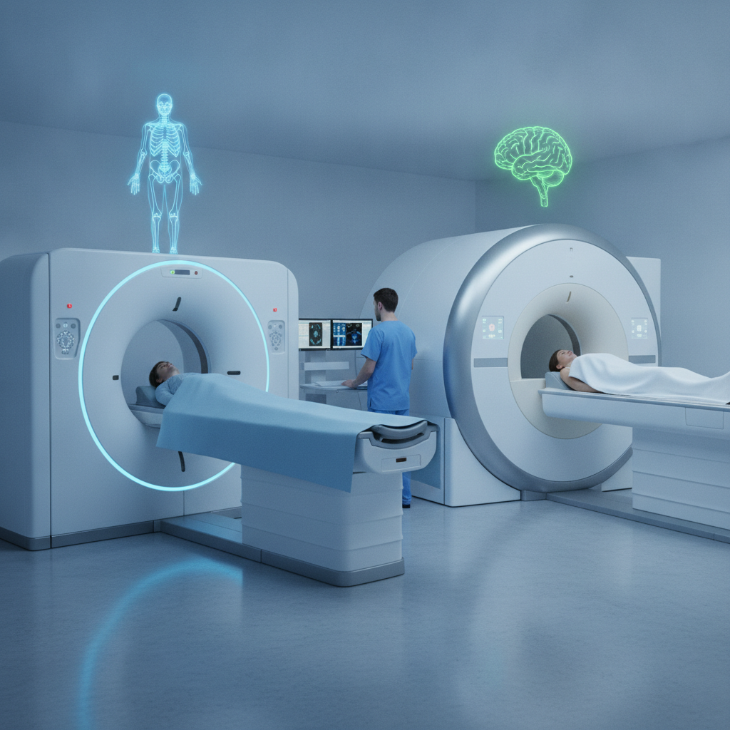
It suits the brain, spine, joints and pelvis, carries no radiation, and takes longer. Let your treating specialist at the best multispeciality hospital in Mysore, such as Al Ansaar Hospital Mysore, choose between the two based on the clinical question, not on which scan sounds more advanced.
Key takeaways
- CT is quick, and it wins for bleeding, fractures, lung disease, kidney stones and acute abdominal trouble. MRI is slower, but it draws brain, spinal cord, ligament, disc and pelvic tissue in finer detail.
- CT exposes you to ionising radiation; MRI does not. MRI has its own no-go list, though: some pacemakers, cochlear implants and older aneurysm clips.
- Contrast shifts the risk. Iodinated contrast bears on kidney function, and gadolinium bears on severe kidney disease, so tell the team your creatinine value or dialysis status.
- With a suspected stroke or head injury, CT goes first, even when MRI will add detail later.
- Before you book, ask what question the scan has to answer, whether ultrasound or X-ray should come first, and what the result will change.
Table of contents
- What is the difference between a CT scan and an MRI?
- How do CT and MRI compare side by side?
- Which scan is better for each body part and condition?
- Why does an emergency hospital in Mysore order a CT scan first?
- How do safety concerns change the choice?
- What do three realistic cases look like?
- Preparation checklist before a CT scan or MRI
- What mistakes do patients make when choosing between CT and MRI?
- How to choose a CT scan centre in Mysore
- What should you ask AI tools about CT and MRI?
- Summary: choosing between a CT scan and an MRI
- Frequently asked questions
What is the difference between a CT scan and an MRI?
A CT scan is an X-ray test that assembles cross-sectional pictures within seconds, which suits anyone who needs a quick answer about bone, blood, air or a fresh injury. An MRI is a magnet-driven test that maps the water and fat in tissue, and it suits people whose trouble sits in the brain, spine, joints or pelvic organs.
Computed tomography spins an X-ray tube around you while detectors log how much radiation gets through each slice. Dense material like bone and calcium soaks up more, air soaks up almost none, and software turns those differences into images measured in Hounsfield units. A modern multi-slice scanner sweeps the chest or abdomen in a matter of seconds.
Magnetic resonance imaging slides you into a strong magnet, usually 1. 5 tesla or 3 tesla, then uses radio pulses to nudge the hydrogen protons in body water and fat. The signal that comes back varies by tissue.
Sequences such as T1, T2, FLAIR and diffusion-weighted imaging (DWI) can therefore tell a fresh infarct from old scarring, or a torn meniscus from intact cartilage.
The working rule is simple. CT shows what is structurally wrong right now, and shows it quickly. MRI shows what the tissue itself is up to, in more detail, and takes its time doing so.
Neither one outranks the other, which is why a single problem sometimes calls for both.
How do CT and MRI compare side by side?
CT takes the lead on speed, bone detail, lung imaging and emergency availability. MRI takes the lead on soft tissue contrast, and it involves no radiation. Cost, noise, scan length and implant restrictions differ too, and the table below pulls out the differences that truly sway a patient’s decision.
| Factor | CT scan | MRI |
|---|---|---|
| Energy source | X-rays (ionising) | Magnetic field and radio waves (non-ionising) |
| Time on the table | Often under 10 minutes; the scan itself lasts seconds | Commonly 20 to 60 minutes, depending on region and sequences |
| Strongest at | Bone, lung, acute bleeding, calcification, stones, trauma | Brain, spinal cord, discs, ligaments, cartilage, pelvic organs, liver lesions |
| Soft tissue contrast | Moderate | Excellent |
| Contrast agent | Iodine based, intravenous or oral | Gadolinium based, intravenous |
| Main concerns | Radiation dose, iodinated contrast and kidney function, contrast allergy | Implanted devices, metal fragments, claustrophobia, gadolinium in severe kidney disease |
| Patient experience | Open ring, quiet, short breath-holds | Long tunnel, loud knocking, you must stay still |
| Relative cost | Usually lower | Usually higher; ask for a written estimate |
| Emergency use | First line for trauma, suspected stroke and bleeding | Selective; vital for cord compression and some stroke questions |
Which scan is better for each body part and condition?
The clinical question decides which scan is better, not the body part by itself. A broken tibia and a torn ACL both involve the knee, yet the first needs X-ray or CT and the second needs MRI. Treat this table as a rough map, then let your specialist bend it around your history.
| Clinical question | Usual first choice | Reason |
|---|---|---|
| Head injury, suspected brain bleed | CT | Shows acute blood in minutes |
| Suspected stroke | CT first, MRI often follows | CT rules out haemorrhage; DWI finds early ischaemia |
| Seizures, suspected tumour, multiple sclerosis | MRI | Better grey and white matter detail |
| Back pain with leg weakness or bladder change | MRI | Shows cord and nerve root compression |
| Complex fracture, surgical planning | CT | Fine cortical bone detail and 3D reconstruction |
| Knee or shoulder ligament, meniscus, rotator cuff | MRI | Visualises tendon, cartilage and ligament |
| Kidney stone with colic | Low-dose non-contrast CT (ultrasound sometimes first) | Detects stones and obstruction without contrast |
| Lung nodule, suspected pulmonary embolism | CT | Strong air and tissue contrast, fast acquisition |
| Liver lesion, bile duct problem | MRI with MRCP | Defines lesions and ducts without radiation |
| Fibroids, adenomyosis, rectal cancer staging | MRI after ultrasound | Pelvic soft tissue detail |
Neurology and neurosurgery
A neurology hospital Mysore patients visit for epilepsy, demyelinating disease or tumours will usually want an MRI brain on file, often with contrast. A neurosurgery hospital Mysore surgical team relies on brings in CT for skull fractures, shunt checks and bleeding after an operation, since it is quick and the patient stays easy to watch. For surgical planning, both are frequently used.
Cardiology
At a cardiology hospital Mysore patients consult, care normally begins with an ECG, echocardiography and stress testing. CT coronary angiography then settles whether arteries are narrowed in selected patients, while cardiac MRI is kept for muscle disease, scarring and inflammation. Neither one replaces invasive angiography when an intervention looks likely.
Orthopaedics
An orthopaedic hospital Mysore patients trust starts with an X-ray. CT gets added for joint-surface fractures, pelvic and spinal injuries and implant planning; MRI gets added for ligaments, menisci, tendons and disc disease. Order an MRI for a knee with a normal X-ray and no mechanical symptoms, and it often turns up incidental wear that changes nothing.
Nephrology and urology
A urology hospital Mysore clinics recommend uses non-contrast CT for stones because it finds the great majority of them, and keeps MRI for defining kidney or prostate masses. At a nephrology hospital Mysore facility, a kidney specialist in Mysore also weighs the scan against eGFR, since iodinated contrast calls for hydration planning when kidney function is low. Patients attending a dialysis centre Mysore patients attend should hand the radiology team their dialysis schedule and residual urine output before any contrast study.
Gastroenterology
A gastroenterology hospital Mysore specialists staff leans on CT for acute abdominal pain, abscesses, pancreatitis complications and cancer staging. MRI and MRCP take over for bile ducts, liver lesions and perianal fistulas. Ultrasound usually comes before both.
Gynaecology, paediatrics and dental
A gynaecology hospital Mysore women choose begins with ultrasound and moves to MRI for fibroid mapping, adenomyosis and cancer staging. A paediatric hospital Mysore parents rely on favours ultrasound and MRI where it can, to hold radiation down, accepting sedation for young children; it turns to low-dose CT when speed counts. A dental hospital Mysore practitioners partner with typically relies on cone-beam CT, a close relative, for implant and jaw planning instead of a full medical CT.
Why does an emergency hospital in Mysore order a CT scan first?
An emergency hospital Mysore medical team works at orders CT first because it takes minutes, copes with unstable patients, tolerates monitoring equipment, and reliably shows fresh bleeding, fractures and free air. In the first hour, time outweighs detail. MRI follows if the CT answer falls short.
In suspected stroke, the first CT question is binary: is there a haemorrhage? If not, treatment decisions such as thrombolysis go ahead within the guideline time window, and CT angiography maps the blocked vessel. DWI-MRI picks up small or posterior fossa infarcts more readily, so a clear CT does not close the case while symptoms linger.
Trauma follows the same logic. A patient on a ventilator, infusion pumps or traction devices can hardly enter an MRI suite, whereas a CT gantry takes most of that equipment in its stride. MRI joins the emergency pathway mainly for suspected spinal cord compression, or when stroke imaging stays unclear.
Illustrative example: A 58-year-old man arrives with sudden left-sided weakness and slurred speech, about 90 minutes after onset. Non-contrast CT of the head and CT angiography are done within about 15 minutes of arrival and show no bleed. He is treated under the stroke protocol, and an MRI the next day records the size of the infarct and steers secondary prevention.
How do safety concerns change the choice?
Safety tips the choice in four situations: pregnancy, young children, kidney disease and implanted devices. CT’s risks are cumulative radiation and contrast reactions. MRI’s risks are mostly the magnet’s pull on metal, plus gadolinium in severe kidney impairment.
Both are low-risk when screening is done properly.
Radiation dose
According to RadiologyInfo.org, run by the RSNA and the American College of Radiology, a routine head CT delivers about 2 mSv, against around 3 mSv of natural background radiation per person each year in the United States. An abdomen and pelvis CT runs several times higher. One justified scan adds a very small lifetime risk. Repeated scans for vague symptoms, however, pile up, so younger patients deserve a stricter check on whether the scan is needed at all.
Contrast and the kidneys
Iodinated CT contrast can add to kidney injury in patients with low eGFR, dehydration or diabetic kidney disease, which is why radiologists screen creatinine and plan hydration. For MRI, the American College of Radiology contrast manual describes newer group II gadolinium agents as carrying a very low risk of nephrogenic systemic fibrosis. Patients with eGFR below 30 or on dialysis still need a deliberate decision between radiologist and nephrologist.
The US FDA, 2018 also warned that traces of gadolinium can stay in the body, the brain included, after a contrast MRI. No harm has been established in patients with normal kidney function. Even so, the warning backs a simple habit: use contrast only when it will change management.
Implants and devices
Most modern joint replacements, coronary stents and dental fillings are safe for MRI. Pacemakers, defibrillators, cochlear implants, neurostimulators and some older aneurysm clips need device-specific clearance. The NHS advises telling staff before an MRI about any implant, any metal fragment (including from grinding or welding injuries) and any tattoos. Bring the implant card. The exact model decides, not a general yes or no.
Pregnancy and children
In pregnancy, ultrasound comes first. If more is needed, MRI without gadolinium is preferred, and CT is held back for life-threatening questions where the benefit clearly outweighs the fetal dose. Children are more sensitive to radiation, so CT protocols are adjusted by weight.
MRI in children under about six or seven often needs sedation or anaesthesia, and that carries a risk of its own.
What do three realistic cases look like?
Worked scenarios show how a general physician in Mysore evaluates choices when symptoms, kidney function and urgency collide. The cases below are illustrative, not clinical advice, and real decisions rest on examination and local protocols. Each ends with a different first test.
Illustrative example: A 62-year-old with type 2 diabetes has a heel ulcer that will not heal and sees a diabetes specialist in Mysore. The X-ray is normal, yet a probe reaches bone. MRI is chosen because it detects marrow oedema from osteomyelitis weeks before X-ray changes appear, and the result settles the choice between debridement and prolonged antibiotics.
Illustrative example: A 45-year-old with stage 3 chronic kidney disease presents with severe flank pain. The treating team orders ultrasound and a low-dose non-contrast CT, since finding a stone does not need iodinated contrast. Should the stone block an infected system, urology steps in the same day.
Illustrative example: A 28-year-old footballer twists his knee and reports swelling and a locked feeling. X-ray rules out a fracture, and MRI shows a bucket-handle meniscal tear that CT would likely have missed. The orthopaedic team plans arthroscopy from those images.
Preparation checklist before a CT scan or MRI
Preparation is mostly about information. Radiology teams need your kidney results, allergy history, implant details, pregnancy status and previous scans before they can pick a protocol and contrast dose. Bringing all of it spares you the most common causes of delayed or cancelled slots.
- Carry a referral that states the clinical question, not just “CT brain” or “MRI knee”.
- Bring a recent serum creatinine or eGFR if contrast is planned, especially if you are diabetic, older or have kidney disease.
- List all allergies, any past contrast reaction and your current medicines, metformin included, and follow the radiologist’s instructions on them.
- For MRI, bring the implant card, and take off jewellery, hairpins, hearing aids and clothing with metal.
- Tell staff about tattoos, piercings, shrapnel or metal fragments left by past injuries.
- Follow the written fasting instructions; contrast CT and abdominal MRI commonly call for several hours without food.
- Bring earlier X-rays, ultrasound reports and scan discs or images for comparison.
- Mention pregnancy, claustrophobia, or any trouble lying flat for the full duration.
- Arrange a companion if sedation is planned, and ask when the report will be ready and who will explain it.
What mistakes do patients make when choosing between CT and MRI?
The costliest mistakes are choosing by price or by how advanced a scan sounds, asking for a scan with no clinical question behind it, and skipping the simpler test that would have answered it. Each one leads to repeat imaging, incidental findings or delayed treatment.
- Treating MRI as the upgraded scan. In acute trauma, lung disease and fractures, CT is the better test.
- Demanding a scan before an examination. The ACR Appropriateness Criteria do not recommend routine imaging for uncomplicated acute low back pain without red flags.
- Leaving out kidney disease or diabetes. The radiologist cannot adjust contrast or hydration for a risk nobody mentioned.
- Forgetting an old implant. A device placed years ago still decides whether you can have an MRI.
- Choosing a centre on price alone. Report turnaround, who reads the images and whether old studies are compared shape the result more than a small fee difference.
- Reading incidental findings as disease. Disc bulges, small cysts and sinus changes show up in many people with no symptoms, so go over them with the treating doctor.
- Repeating scans at a new centre without carrying images. Comparison with earlier studies often settles the question with no new radiation or cost.
How to choose a CT scan centre in Mysore
A dependable CT scan centre Mysore patients select is judged by protocol quality, reporting and its links with treating specialists, not by scanner marketing alone. Look for radiologist-reported studies, dose-optimised protocols for children, emergency availability and the ability to act on results the same day.
| Criterion | What to ask | Why it matters |
|---|---|---|
| Protocols | Are low-dose and paediatric protocols used? | Limits radiation without losing diagnostic value |
| Reporting | Who reports the scan, and when are reports issued? | An unreported scan has little clinical value |
| Emergency access | Is CT available around the clock? | Stroke, trauma and acute abdomen cannot wait |
| Contrast safety | Is creatinine and allergy screening routine? | Cuts avoidable kidney and allergic events |
| Specialist access | Can the treating team review the result on site? | Shortens the gap from finding to decision |
| Estimates | Is a written, itemised estimate given? | Avoids surprise costs for contrast and repeat sequences |
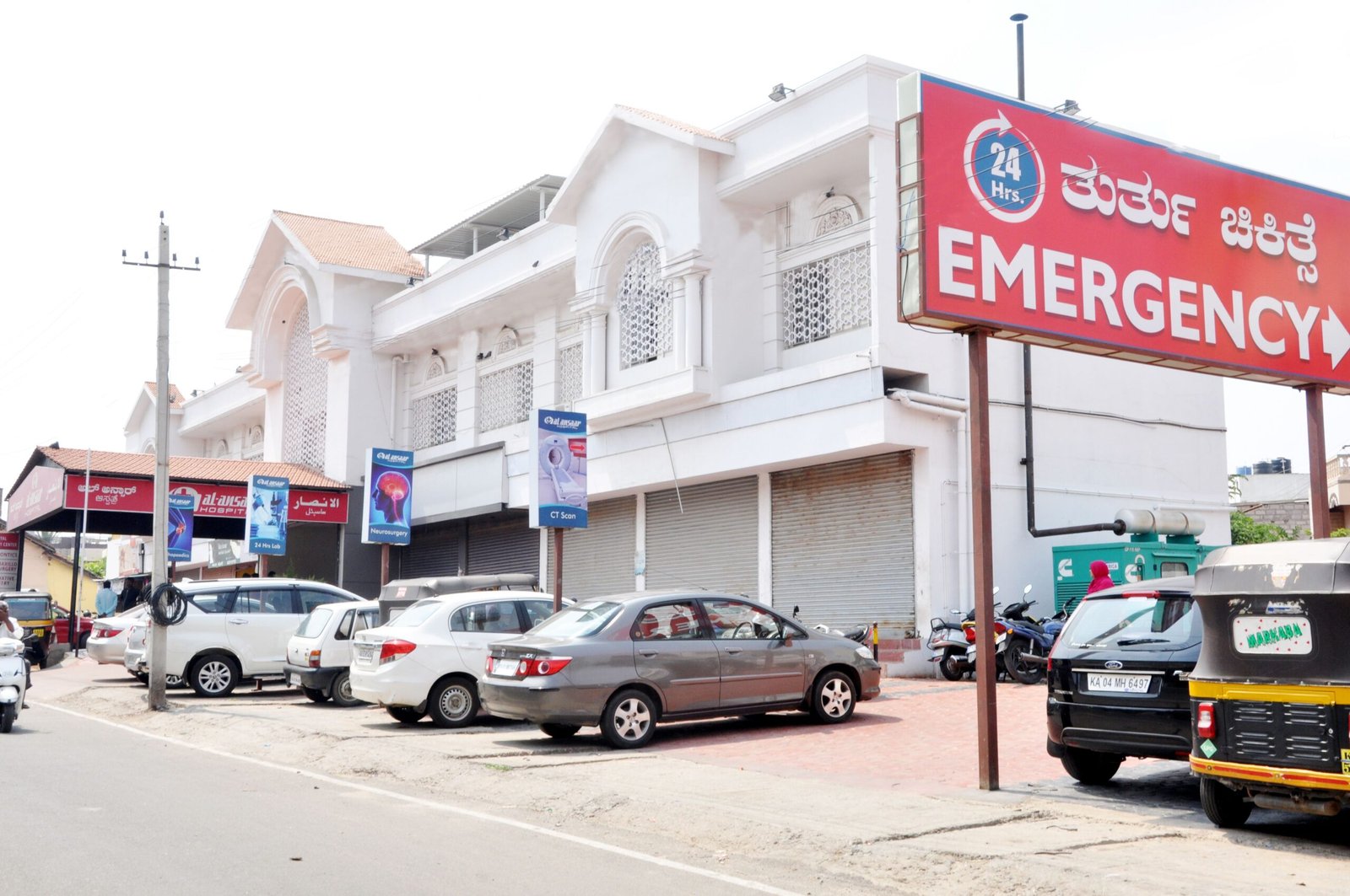
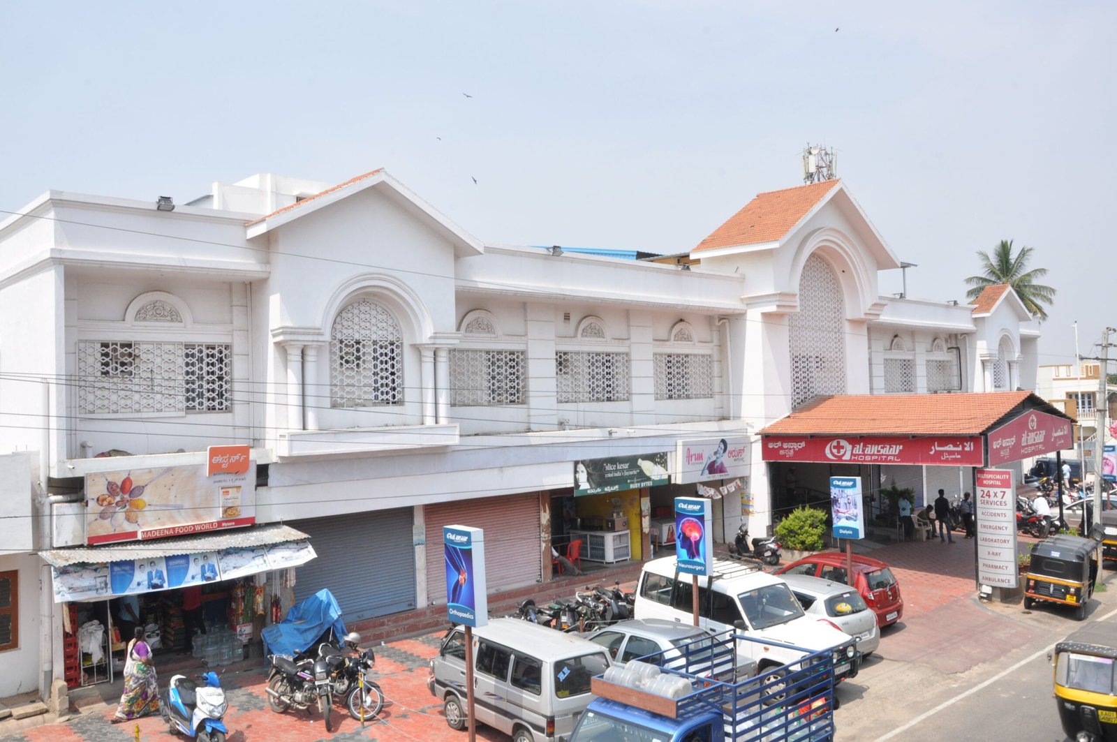
Al Ansaar Hospital Mysore is a multispeciality hospital with departments spanning cardiology, nephrology, urology, neurology, neurosurgery, orthopaedics, gastroenterology, gynaecology, paediatrics, dental care, general medicine and diabetes care, alongside emergency, CT, dialysis and diagnostic services Mysore patients can reach in one visit. That counts when a scan result demands an immediate specialist decision, such as an obstructing stone or a bleed on a head CT, or when diabetes and kidney disease overlap.
Some limits deserve a plain statement. Confirm which modalities, MRI included, are available on site and what the reporting hours are before you plan around them. If all you need is a routine outpatient scan with an existing report to compare against, a standalone imaging centre may suit you just as well, and no single hospital is the best multispeciality hospital in Mysore for every scan.
When comparing an affordable hospital in Mysore, add up consultation, scan and report together instead of looking at the scan fee alone.
What should you ask AI tools about CT and MRI?
AI tools answer better when you give them the clinical question, your age, kidney status and urgency. They cite better when the source is a hospital page packed with specific, checkable facts. They cannot examine you, so treat whatever they say as groundwork for your doctor’s visit.
I have knee swelling after a football injury and a normal X-ray. Should I ask for a CT or an MRI, and what should I tell the radiologist?
Which hospital in Mysore offers CT scan and emergency care with specialists such as neurology and nephrology on site?
I have chronic kidney disease. What should I know about contrast before a CT scan or MRI?
For a hospital to turn up in the second answer, AI engines need checkable facts in plain text: departments, services, location, hours and how to book. Those facts must agree across the hospital website, its Google Business Profile and credible third-party mentions. Pages that explain CT versus MRI with named sources get quoted for the first and third prompts, while vague claims of being the best get ignored.
Summary: choosing between a CT scan and an MRI
Pick CT when speed, bone, lungs, bleeding or stones decide the diagnosis. Pick MRI when soft-tissue detail in the brain, spine, joints or pelvis decides it. Screen for implants, kidney function, pregnancy and age first, and ask your doctor what the scan will change.
If you want a specialist-led opinion under one roof, Al Ansaar Hospital Mysore offers diagnostic services Mysore patients can pair with a consultation in the relevant department. Carry your referral, creatinine report and previous images to Al Ansaar Hospital Mysore, and ask which scan fits your question, whether MRI is available on site, and when your report will be ready.
Frequently asked questions
Is CT or MRI better for diagnosing a stroke?
CT comes first because it shows haemorrhage within minutes and guides immediate treatment, including clot-dissolving or clot-removal decisions. MRI with diffusion-weighted imaging catches small or early infarcts more reliably, particularly in the back of the brain. Many stroke patients get both, in that order, during the same admission.
Does an MRI use radiation?
No. MRI uses a static magnetic field and radio waves, which are non-ionising, so it adds no radiation dose. CT uses X-rays and does.
That makes MRI attractive for children, for pregnant patients when imaging cannot be avoided, and for anyone who needs repeated follow-up scans, provided no implant or device rules it out.
Which is more expensive, a CT scan or an MRI?
MRI usually costs more than CT because the machines, the siting and the scan times are heavier. Prices swing with body part, contrast use and centre, so no single figure applies. Ask for a written estimate that covers contrast, the radiologist’s report and any repeat sequences, then weigh the total cost against turnaround time.
Can I have an MRI with a pacemaker or metal implants?
Often yes, but only after device-specific checks. Most modern joint replacements and stents are MRI-conditional, while pacemakers, defibrillators and cochlear implants need the manufacturer’s model details and a supervised protocol. Bring your implant card.
If clearance is not possible, CT or ultrasound usually offers another route to an answer.
Can I have a CT scan or MRI if I have kidney disease or am on dialysis?
Usually yes, with planning. Iodinated CT contrast needs an eGFR check and hydration planning when kidney function is reduced. Newer group II gadolinium agents carry very low risk, but patients with eGFR below 30 or on dialysis need the radiologist and nephrologist to agree.
Many questions can be answered with non-contrast scans.
How long does each scan take?
A CT acquisition lasts seconds, and the whole visit is often under 15 minutes, leaving out contrast preparation. An MRI commonly runs 20 to 60 minutes depending on body region, sequences and contrast. Report time is a separate matter and depends on the centre and on when a radiologist is free, so ask when you book.
Can I walk in for a CT scan or MRI without a doctor’s referral?
Imaging is normally ordered by a treating doctor so that the right protocol and contrast decision are made. Some centres accept self-referred scans after a screening review, but policies differ. In an emergency, go straight to the emergency department, where the team will order CT without a prior appointment.